Nobel Prize Winner Optogenetics: How Light Controls Brain Cells and Could Transform Medicine
Nobel Prize Winner Optogenetics. Imagine a future in which scientists can activate or silence selected nerve cells simply by delivering a carefully controlled beam of light. It sounds like science fiction, yet the underlying technology is already transforming neuroscience research.
https://mrpo.pk/tesla-roadster-2026/
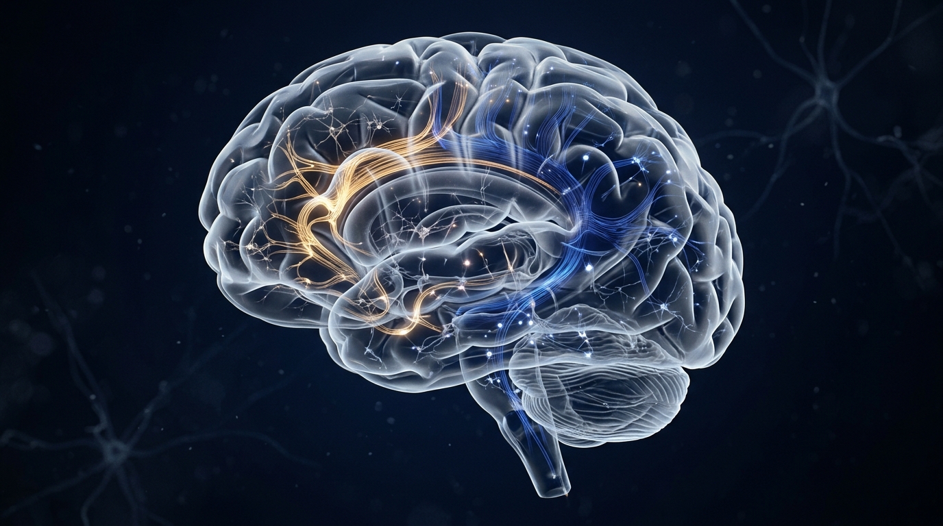
Known as optogenetics, this breakthrough was recognised by the 2026 Nobel Prize in Physiology or Medicine, awarded to Karl Deisseroth of the United States and Peter Hegemann and Georg Nagel of Germany for their discoveries concerning light-gated ion channels and optogenetics. The award was announced on 5 October 2026. Official Nobel Prize announcement.

Credit: Cmichel67/Wikimedia Commons (Karl Deisseroth); Associated Press (Peter Hegemann); The Brain Prize (Georg Nagel)
Their discoveries have given researchers a remarkable way to investigate how the brain works, from the electrical activity of individual nerve cells to the complex networks involved in movement, memory and behaviour. The three laureates share a prize of 12 million Swedish kronor, in addition to receiving Nobel medals and diplomas.
But how can light influence a living brain? Why did the discovery begin with microscopic green algae? Could this technology eventually help people with epilepsy, chronic pain, neurological disorders, or blindness? And how far are we from using it routinely in hospitals?
This article explains the science from beginning to end in simple language, while distinguishing established research capabilities from treatments that are still being investigated.
What Is Optogenetics?
Optogenetics combines two scientific fields: genetics, which studies genes and biological instructions, and optics, which deals with light.
To understand its purpose, imagine the brain as an enormous communication network containing billions of nerve cells, known as neurons.
These neurons communicate through electrical impulses and chemical messengers. Their coordinated activity allows us to move, learn, remember, recognise faces, experience emotions and respond to our surroundings.
Scientists have long sought to understand exactly how particular groups of neurons contribute to these functions.
Traditional approaches, including medication and electrical stimulation, can influence nerve activity, but they may affect multiple cells or pathways simultaneously. Medicines can also affect other organs, while electrical stimulation may activate neighbouring nerve fibres unintentionally.
Optogenetics offers a more selective research method. Scientists make particular cells sensitive to light and then use controlled illumination to influence their electrical activity.
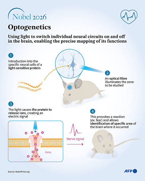
The essential idea is simple: give selected cells a light-sensitive biological switch, activate that switch with light, and observe what changes.
This is not the same as shining a flashlight at someone’s head and controlling their thoughts. The target cells must first be made light-sensitive, and specialised equipment is generally required to deliver light to the appropriate tissue.
The Discovery: How Pond Algae Helped Unlock the Brain
One of the most fascinating aspects of optogenetics is that its foundations came from studying a microscopic organism rather than a human brain.
Nature’s Light-Sensitive Organism
Researchers investigating the single-celled green alga Chlamydomonas wanted to understand how it detects and responds to light.
Light helps this organism navigate towards favourable conditions for photosynthesis. Scientists discovered specialised proteins that enable it to respond to illumination.
Among these proteins were channelrhodopsins, light-sensitive proteins that form channels in cell membranes.
Think of a cell as a tiny room surrounded by a wall. That wall regulates the movement of electrically charged particles into and out of the cell. Channelrhodopsin acts like a molecular door that opens in response to the appropriate light.
When the door opens, ions can cross the membrane, changing the cell’s electrical state.
From Algae to Nerve Cells
Peter Hegemann and Georg Nagel made foundational contributions to understanding microbial light-sensitive proteins, including channelrhodopsins. Karl Deisseroth and other researchers helped turn these discoveries into a powerful method for controlling neurons.
The crucial insight was that the genetic instructions for these proteins could be introduced into selected animal nerve cells.
Those cells could then produce light-sensitive proteins in their membranes. Researchers could activate the proteins using appropriate light and thereby influence the cells’ electrical activity.
What began as an investigation into how tiny algae respond to sunlight became a new way to investigate the living brain.
This is an important lesson about scientific progress: discoveries that initially appear to have little immediate medical value can eventually transform an entire field.
How Light Controls Brain Cells: The Complete Process from A to Z
To understand optogenetics properly, it helps to follow the entire process in the order it happens.
Imagine that researchers want to investigate a particular group of neurons involved in epileptic seizures. They want to determine whether changing the activity of those cells can reduce abnormal electrical activity elsewhere in the brain. The process involves several connected stages.
These proteins became the foundation of optogenetics, the technology we have been discussing.
1. How do these proteins work?

Consider a tiny green alga called Chlamydomonas. It consists of just one cell, yet it can detect light and adjust its swimming direction to find favourable conditions for photosynthesis.
Here is how a light-sensitive protein works:

Step 1: Light arrives
Light consists of tiny packets of energy called photons. A light-sensitive part of the protein absorbs a photon of the appropriate wavelength.

Step 2: The protein changes
Absorbing light changes the shape or state of the protein, triggering a biological response.

Step 3: The cell responds
In proteins such as channelrhodopsin, the change opens an ion channel. Electrically charged particles cross the cell membrane, changing the cell’s electrical state.
2. What is channelrhodopsin?

Channelrhodopsin is a light-sensitive protein originally identified in green algae. It acts like a tiny molecular gate in the cell membrane.
When suitable blue light activates channelrhodopsin, the gate opens and allows certain ions to pass through. This changes the electrical voltage across the membrane.
In algae, this contributes to their ability to detect and respond to light. Scientists realised that the same protein could also be used to influence electrical activity in nerve cells.
That was a major breakthrough.
3. How did an algal protein become a tool for studying the human brain?
Scientists introduced the genetic instructions for channelrhodopsin into selected neurons in laboratory experiments.
The modified neurons then produced the light-sensitive protein in their membranes.
When researchers delivered the appropriate light, the protein opened its channel, ions moved across the membrane, and the neuron could generate electrical impulses.
This allowed scientists to investigate what happens when particular brain cells are activated at a chosen moment.
An important distinction: the protein does not create a complete electrical signal by itself. It changes the cell’s electrical conditions so that the neuron’s own biological machinery can generate or modify its activity.
4. Are all light-sensitive proteins the same?
No. Different proteins respond to light in different ways.
| Protein type | What it does |
|---|---|
| Channelrhodopsins | Open channels that allow ions to cross a cell membrane |
| Light-driven ion pumps | Move particular ions across a membrane using light energy |
| Other opsins | Trigger different cellular responses, depending on their structure and biological context |
Researchers choose a protein according to whether they want to stimulate a cell, inhibit its activity or investigate another light-dependent process.
The big idea
Imagine installing a light-operated switch in a living cell. The switch is made from a protein, the instructions for producing it come from a gene, and light activates it.
That is the basic principle behind optogenetics.
What makes the discovery extraordinary is that scientists borrowed a mechanism from a microscopic organism and adapted it to investigate one of the most complex structures in nature: the human brain.
Step 1: Identify the Target Cells
First, scientists identify the particular neurons or neural pathways they want to investigate.
These might be cells involved in movement pathways, memory, sensory perception or seizure activity.
The selection is important because different neurons perform different functions. Some excite other neurons, increasing the likelihood that they will fire. Others inhibit their activity.
Researchers use specialised genetic markers, anatomical information and other laboratory methods to identify the relevant cell populations.
Step 2: Introduce the Genetic Instructions
The selected cells must be made responsive to light.
Scientists introduce genetic instructions that enable those cells to manufacture a suitable light-sensitive protein, known as an opsin.
One commonly used example is channelrhodopsin-2, or ChR2.
In experimental research, engineered viral vectors are often used to deliver these instructions. The vectors act as delivery vehicles for genetic material; they do not themselves switch the neurons on or off.
Other delivery methods may be suitable depending on the experimental system.
The objective is to make the intended cells produce the protein while limiting unintended expression in other cells.
Step 3: The Cells Manufacture the Light-Sensitive Protein
Once the genetic instructions reach the appropriate cells and are expressed, the cells begin producing the protein.
The protein is transported to the cell membrane, where it can interact with light.
The cell remains alive and continues to perform its normal biological functions, but it now has an additional mechanism for responding to illumination.
An everyday analogy would be installing a specialised light-sensitive switch in the wall of a room. The switch is part of the system, but it needs the correct signal before it changes the system’s behaviour.
Step 4: Deliver the Appropriate Light
Researchers now deliver controlled light to the target tissue.
Depending on the experiment, they may use LEDs, lasers, optical fibres, or other specialised optical equipment.
The light must have an appropriate wavelength for the selected protein. Its intensity, timing and duration must also be carefully controlled.
For example, blue light can activate ChR2. Other light-sensitive proteins respond to different wavelengths or produce different cellular effects.
For neurons located deep within the brain, an optical fibre may be positioned close to the target tissue. Other arrangements are possible for cells closer to the surface or in accessible tissues.
This is one reason optogenetics is not equivalent to shining ordinary light at someone’s head. Light scatters and is absorbed as it travels through biological tissue, making precise delivery difficult.
Step 5: Light Changes the Shape of the Protein
When an appropriate photon of light is absorbed by a light-sensitive protein, it triggers a molecular change.
In channelrhodopsins, this change opens a channel through which certain electrically charged particles can pass across the cell membrane.
The protein therefore converts a physical signal, light, into a biological response.
This molecular event is the heart of the technology.
Step 6: Charged Particles Change the Cell’s Electrical State
Neurons maintain an electrical voltage across their membranes. This voltage depends on the distribution of ions, including sodium, potassium and chloride, and on the properties of the cell membrane.
When ChR2 opens, positively charged ions can enter the neuron, changing the voltage across its membrane. The inside becomes less electrically negative, a process called depolarisation.
If the voltage change reaches the necessary threshold, the neuron can generate an action potential, an electrical impulse that travels along the cell.
In simple language, light activates the protein, the protein permits ions to move, and that movement can cause the nerve cell to fire.
Light does not manufacture an electrical impulse by itself. It triggers a molecular mechanism that interacts with the cell’s existing electrical machinery.
Step 7: Researchers Can Activate or Inhibit Neurons
Optogenetics is not limited to switching cells on.
Different light-sensitive proteins can produce different effects. Some encourage neurons to fire; others can reduce their likelihood of firing.
Certain inhibitory opsins move ions across the membrane using light-driven pumps, while others alter the membrane voltage through different mechanisms.
The effect depends on the protein, the wavelength of light, the cell type and the experimental conditions.
This distinction is particularly important when researchers investigate epilepsy. They may want to activate inhibitory neurons, suppress particular overactive cells or investigate how different combinations affect the wider neural network.
Step 8: The Neurons Influence Other Cells
A neuron does not work in isolation.
When it fires, its electrical signal can travel along its axon towards connections called synapses. At many synapses, this triggers the release of neurotransmitters, chemical messengers that influence neighbouring cells.
Some neurotransmitters increase the likelihood that a receiving neuron will fire. Others reduce it.
Consequently, changing the activity of one selected group of neurons can influence an entire neural circuit.
Researchers use this capability to investigate how individual cells contribute to more complex functions, such as movement, learning, memory and sensory processing.
However, activating one group of neurons does not guarantee a particular behaviour. The outcome depends on the cells involved, their connections, the timing of stimulation and the wider state of the brain.
Step 9: Measure the Results
Once the cells have been stimulated, researchers investigate what happens.
They may use electrical recording equipment to measure nerve impulses, imaging methods to observe cellular activity or behavioural experiments to examine changes in an experimental animal.
For example, if activating a particular neural pathway repeatedly changes a movement, researchers can investigate whether that pathway contributes to controlling the movement.
They can then compare the results with appropriate controls and test whether inhibiting the same pathway changes the outcome.
This is one of optogenetics’ greatest strengths: scientists can manipulate a selected biological component and investigate cause and effect, rather than simply observing that two events occur together.
Step 10: Switch Off the Light and Continue the Experiment
When illumination stops, the light-sensitive protein generally returns to its inactive state according to its molecular properties.
The etoiminishes, although the exact timing depends on the protein. A neuron may continue firing because of other signals it is receiving, and downstream changes may persist after the light is removed.
Researchers can adjust the timing and pattern of stimulation, repeat experiments and compare different conditions.
Optogenetics does not reset the whole brain. It gives scientists a controllable way to influence selected cells and examine the consequences.
The Entire Process at a Glance
The complete sequence can be summarised as follows:
- Identify: Select the neurons or neural pathway to investigate.
- Introduce: Deliver genetic instructions for a suitable light-sensitive protein.
- Produce: Allow the selected cells to manufacture the protein.
- Illuminate: Deliver controlled light to the target tissue.
- Activate: The protein changes state and alters ion movement.
- Influence: The change can increase or reduce neuronal firing.
- Measure: Record electrical activity, cellular responses or behaviour.
- Interpret: Compare results to understand the function of the neural circuit.
This combination of genetic targeting, light-sensitive proteins and precise illumination is what makes optogenetics so powerful.
Why Optogenetics Is a Scientific Breakthrough
The brain is a highly interconnected system. Different groups of neurons cooperate to perform many functions, and the same region can participate in several processes.
Before optogenetics, scientists had limited ways to manipulate specific types of neurons with precise timing while observing the results.
Optogenetics helps answer questions such as:
- Which neural circuits contribute to forming or retrieving memories?
- How do particular pathways influence movement and sensory perception?
- Which cell populations contribute to abnormal activity during a seizure?
- How do specific neural circuits influence motivation, stress and behaviour?
- Which cellular mechanisms might be targeted to develop more selective treatments?
The technique has transformed neuroscience because it allows researchers to test how particular cells contribute to a process, rather than merely observing which cells become active during it.
Nevertheless, it does not mean that complex experiences such as consciousness, love or sadness can be reduced to a single switch. Such experiences involve multiple interacting brain networks, bodily processes and environmental influences.
Potential Medical Applications: From Seizures to Chronic Pain
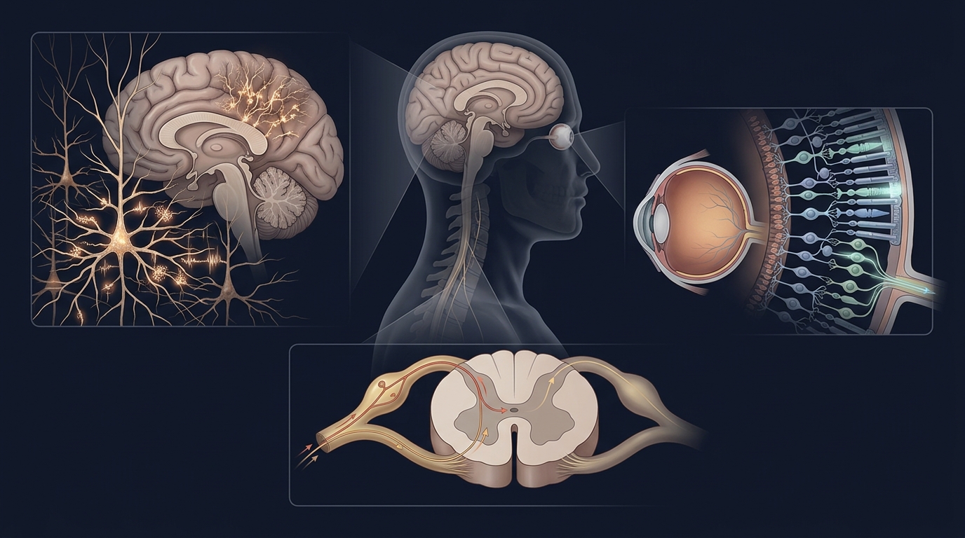
Optogenetics is already an established research tool, but many of its possible medical applications remain experimental. Its value lies both in investigating disease mechanisms and in exploring whether light-sensitive cells could eventually form part of new treatments.
Epilepsy and Seizures
Epilepsy involves a tendency to experience recurrent seizures caused by abnormal electrical activity in the brain.
Researchers use optogenetics to investigate whether activating inhibitory neurons or suppressing selected overactive cells can reduce seizure activity in experimental models.
One possible future approach is a closed-loop system that detects signs of an approaching seizure and delivers an intervention when needed. An optogenetic version would require light-sensitive cells and a specialised system for delivering light to the appropriate brain tissue.
However, significant challenges remain, including safe gene delivery, accurate targeting and reliable long-term operation.
Optogenetics is not currently a routine treatment for epilepsy. Its major contribution is helping scientists understand seizure mechanisms and investigate potential therapeutic approaches.
Chronic Pain and Spinal Nerve Disorders
Persistent pain can develop when damaged nerves or spinal pathways become unusually sensitive. In some cases, pain continues even after the original injury has healed.
Researchers can use optogenetics in experimental models to activate or inhibit selected nerve cells and investigate how pain signals travel from peripheral nerves through the spinal cord to the brain.
This research may help identify pathways that amplify pain and others that suppress it, potentially guiding the development of more selective treatments.
However, optogenetics does not repair a herniated disc, reverse spinal compression or currently provide an established cure for sciatica or chronic back pain. Its principal role in these areas remains experimental research.
Depression, Anxiety and Other Complex Conditions
Researchers also investigate brain circuits involved in mood, motivation, stress and behaviour.
By manipulating selected neural populations in experimental models, scientists can investigate how particular circuits contribute to certain responses and how changes in these circuits might relate to psychiatric conditions.
Similar research may help improve understanding of chronic fatigue syndrome, also known as myalgic encephalomyelitis or ME/CFS, a complex illness whose underlying mechanisms remain incompletely understood.
These conditions cannot be explained by a single brain circuit or a simple biological switch. Optogenetics is a research tool that may help clarify some of the mechanisms involved; it is not an established diagnostic test or treatment for depression, anxiety, or ME/CFS.
Could Optogenetics Restore Vision?
Vision restoration is a distinct area of clinical investigation.
In certain retinal diseases, the cells that normally detect light become damaged or die, while some other retinal cells may survive.
Researchers are investigating whether introducing genes for light-sensitive proteins into surviving cells can help restore some ability to detect visual information.
In this approach, the aim is to make surviving retinal cells responsive to light even when the original photoreceptors have been lost. Depending on the technique, specialised optical equipment may be needed to provide an appropriate visual stimulus.
Research has progressed into human clinical investigation, including studies involving people with severe retinal degeneration. However, the amount and quality of restored vision can vary, and the approach does not reproduce normal vision in every patient.
Optogenetic vision restoration is therefore a specialised investigational treatment rather than a universal cure for blindness.
Is Optogenetics Already Being Used in Human Medicine?
The answer depends on whether we are talking about research or routine treatment.
Optogenetics is widely used in laboratories to investigate neural circuits, cellular activity and disease mechanisms. Researchers also use insights from optogenetic experiments to guide the development of treatments that may not themselves involve light-sensitive proteins.
Direct optogenetic interventions in humans are much more limited and remain specialised. Vision restoration is one area where clinical investigation has progressed significantly. The distinction matters because a successful laboratory experiment does not automatically translate into an effective treatment.
Before a new therapy becomes routine, researchers must establish its safety, effectiveness, durability and practical advantages over existing treatments. They must also address regulatory requirements, manufacturing challenges and long-term monitoring.
A Nobel Prize recognises an important scientific achievement; it does not mean every potential application is already available to patients.
Global Research and International Collaboration
Optogenetics is an international scientific endeavour, involving neuroscience, genetics, optical engineering, biomedical technology and clinical research.
The United States has made major contributions through university laboratories and neuroscience research programmes. Karl Deisseroth’s work at Stanford University helped establish optogenetics as a powerful method for studying neural circuits.
Germany played a foundational role through the research of Peter Hegemann and Georg Nagel, whose investigations into light-sensitive proteins in microorganisms helped make the technology possible.
Research groups throughout Europe and Asia are also investigating new light-sensitive proteins, improved gene-delivery methods, optical systems and possible therapeutic applications.
Progress depends on collaboration among biologists, neurologists, geneticists, engineers and clinicians. Advanced microscopes, precision optical equipment, electrical recording systems and sophisticated data analysis all contribute to the field.
For countries such as Pakistan, participation need not begin with the most expensive clinical equipment. Universities and medical research institutions can build expertise through neuroscience education, biomedical engineering, international collaborations and shared research facilities.
The broader lesson is that scientific capability grows through sustained investment in people, knowledge and research infrastructure.
How Expensive Is Optogenetics?
The cost varies considerably according to the purpose of the work.
Research Laboratory Costs
A laboratory investigating optogenetics may need controlled light sources, optical fibres, specialist electronics, recording equipment, microscopes and tools for delivering genetic material.
A basic experimental setup may cost several thousand dollars, while advanced systems can cost tens of thousands of dollars or substantially more. These are broad illustrative ranges, not fixed prices; the actual cost depends on the equipment, laboratory requirements and experimental design.
Ongoing expenses may include consumables, specialised biological materials, animal research facilities and staff training.
Potential Clinical Treatment Costs
Human applications can involve additional expenses associated with gene delivery, specialised medical devices, manufacturing, clinical trials, regulatory approval and long-term safety monitoring.
Some advanced biological therapies are expensive, but it would be misleading to assign a single price to future optogenetic treatments before their clinical use and delivery requirements are established.
Their eventual cost will depend on the specific condition, treatment design, manufacturing process, healthcare system and availability of alternatives.
For developing countries, the priority should be to invest strategically in research capacity, training and collaboration rather than assume that every emerging technology must immediately be adopted as a costly clinical service.
The 2026 Nobel Prize: Recognising a New Way to Study the Brain
The 2026 Nobel Prize in Physiology or Medicine recognised Karl Deisseroth, Peter Hegemann and Georg Nagel for their discoveries concerning light-gated ion channels and optogenetics.
The three laureates share a prize of 12 million Swedish kronor, in addition to receiving Nobel medals and diplomas.
Their recognition reflects a fundamental advance in how scientists can investigate living neural systems.
Hegemann and Nagel helped establish the biological foundations through their work on light-sensitive proteins. Deisseroth and other researchers helped develop the methods that enabled scientists to control neuronal activity with light.
The achievement illustrates why fundamental scientific research matters. Studying a microscopic organism led to a technology that now helps researchers investigate the complex workings of the brain.
Scientific discoveries do not always produce immediate treatments. Sometimes their first contribution is to provide a new way of asking questions that were previously difficult or impossible to answer.
What Does the Future Hold?
Optogenetics has already transformed neuroscience research, but its future medical applications will depend on solving several challenges.
Scientists need reliable ways to deliver genetic instructions to the right cells, ensure that light-sensitive proteins function safely over time, deliver light effectively to the target tissue and demonstrate meaningful benefits for patients.
In some conditions, optogenetics may eventually provide a direct therapeutic approach. In others, its most important contribution may be indirect: revealing which cells and pathways should be targeted by medicines, electrical stimulation, or other treatments.
A recent review in Nature Neuroscience discusses both direct optogenetic interventions and the wider therapeutic opportunities arising from knowledge gained through optogenetic research. Read the review on translating optogenetics into human therapies.
The future will therefore depend not only on controlling cells with light, but on translating the resulting knowledge into safe, effective and affordable medical care.
Conclusion: When Light Becomes a Tool for Understanding Life
Optogenetics represents a remarkable meeting of biology, genetics and optical science.
By enabling scientists to influence selected cells with light, it has opened new ways to investigate how neural circuits contribute to movement, memory, perception and behaviour. It is also helping researchers explore potential approaches to neurological disorders and certain forms of vision loss.
Yet its promise must be balanced with scientific discipline. Many possible treatments remain experimental, and translating laboratory discoveries into routine medical care can take years of further research.
The journey from microscopic algae to the mysteries of the human brain reminds us that nature often holds unexpected answers to humanity’s most difficult questions.
The greatest achievement of optogenetics may not simply be the ability to switch selected nerve cells on or off, but the opportunity to understand the intricate biological systems that make human thought, perception and life possible.
For ordinary people, the central lesson is simple: scientists can make selected cells sensitive to light, use that light to change cellular activity and observe how the change affects a wider biological system. This is how an observation about a tiny organism became a powerful tool for exploring the human brain.
Frequently Asked Questions (FAQs)
1. What is optogenetics in simple language?
Optogenetics is a scientific technique that combines genetics with light-sensitive proteins to control selected cells. Researchers use it to activate or inhibit particular nerve cells and investigate how they influence brain functions.
2. How does light actually activate a neuron?
When the appropriate light reaches a light-sensitive protein such as channelrhodopsin, the protein changes shape and opens an ion channel. Charged particles cross the cell membrane, changing its electrical voltage. If the change is sufficient, the neuron generates an electrical impulse.
3. Can optogenetics switch brain cells off as well as on?
Yes. Different light-sensitive proteins can increase or reduce neuronal activity. The effect depends on the protein, the light used and the properties of the targeted cells.
4. Why did the discovery begin with algae?
Researchers studying Chlamydomonas discovered light-sensitive proteins that could respond to illumination. Scientists subsequently adapted these proteins for use in nerve cells, creating a new way to investigate neural circuits.
5. Can scientists control a person’s brain with a flashlight?
No. Optogenetics generally requires selected cells to produce light-sensitive proteins and specialised equipment to deliver appropriately controlled light. Shining an ordinary flashlight at someone’s head does not provide this capability.
6. Can optogenetics cure epilepsy or chronic back pain?
Not at present. Researchers are investigating its potential to understand seizures and pain pathways, but it is not a routine treatment for epilepsy, sciatica or chronic back pain.
7. Can optogenetics restore sight to blind people?
Clinical research is investigating whether making surviving retinal cells light-sensitive can restore some visual function in selected retinal diseases. Results vary, and the approach is not a universal cure for blindness.
8. Is optogenetics available in hospitals?
Most applications remain research-based or investigational. Direct clinical applications are limited, and their availability depends on the particular condition, treatment and supporting clinical evidence.
9. What did the 2026 Nobel Prize recognise?
The Nobel Prize in Physiology or Medicine recognised Karl Deisseroth, Peter Hegemann and Georg Nagel for their discoveries concerning light-gated ion channels and optogenetics.
10. Why is optogenetics important for ordinary people?
It helps scientists investigate how the brain works and understand the biological mechanisms behind disease. This knowledge may eventually support the development of better treatments for neurological disorders and other conditions.
References and Further Reading
- Nobel Assembly at Karolinska Institutet. The 2026 Nobel Prize in Physiology or Medicine: Official announcement.
- Deisseroth, K. Optogenetics. Nature Methods.
- Fenno, L., Yizhar, O. and Deisseroth, K. The Development and Application of Optogenetics. Annual Review of Neuroscience.
- Yizhar, O. and colleagues. Roadmap for direct and indirect translation of optogenetics into discoveries and therapies for humans. Nature Neuroscience, 2025.
- Lindner, M. and colleagues. Optogenetics for visual restoration: From proof of principle to translational challenges. Progress in Retinal and Eye Research, 2022.